Interested in learning
more about LDF?
Welcome

to the Lateral Distal Femur (LDF) DXA website. The purpose of this site is to serve as the resource for anyone interested in learning more about the LDF as a site to measure by DXA for assessment of bone density. On these pages you can learn about the history of the LDF, how to acquire and analyze the LDF, what good scans look like, technical issues of the scanning technique, reporting results and clinical applications.
Many pediatric facilities are routinely using the LDF as a part of their clinical scanning, particularly those centers serving children with physical disabilities; others use the LDF for research purposes only. For either application, the LDF DXA is easily obtained on most patients.
Why the LDF?
Measuring and interpreting the bone density of children presents the clinician with challenges because children’s bones are small and they are growing. This presents particular technical challenges in both the accuracy of measurement and interpretation of results. Official positions on measuring bone density for both adults and children can be found at the website of the International Society for Clinical Densitometry.
Measuring the bone density of people with physical disabilities is especially challenging due to commonly encountered issues with this population which can invalidate the accuracy of DXA results at body sites typically measured (total body, lumbar spine, and proximal femur/hip).
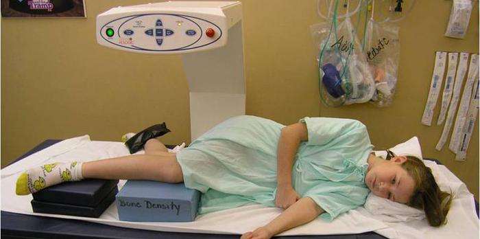
Measuring the distal femur in the lateral projection allows technologists to easily obtain a DXA scan on people with disabilities because it is quick, obtained in a comfortable position, and is less susceptible to movement than traditional body sites measured by DXA.
As with any DXA measurement, scan quality is only as good as the acquisition and analysis. To accurately and consistently perform the LDF DXA, the technologist acquiring and analyzing the scan needs the following skills:
- knowledge of what is being measured
- familiarity with patient positioning and how to make adjustments in positioning to yield the proper view
- familiarity with the DXA scanner and its operation
- knowledge of LDF analysis instructions
The interpreting physician needs to understand:
- what is being measured – types of bone and regions
- what a high-quality scan looks like
- technical considerations for interpretation
- how to report results
This website is for both technologists and clinicians. Browse the pages on this website to learn more about the LDF. In addition to instructions, you will find helpful links, documents and images.
.....................................................